Home » Without Label » Anatomy Of Chest : Diagram Illustrating The Male Chest With Its Associated Arteries Download Scientific Diagram : The thorax or chest is a part of the anatomy of humans, mammals, other tetrapod animals located between the neck and the abdomen.
Anatomy Of Chest : Diagram Illustrating The Male Chest With Its Associated Arteries Download Scientific Diagram : The thorax or chest is a part of the anatomy of humans, mammals, other tetrapod animals located between the neck and the abdomen.
Anatomy Of Chest : Diagram Illustrating The Male Chest With Its Associated Arteries Download Scientific Diagram : The thorax or chest is a part of the anatomy of humans, mammals, other tetrapod animals located between the neck and the abdomen.. The human thorax includes the thoracic cavity and the thoracic wall. The chest wall is comprised of skin, fat, muscles, and the thoracic skeleton. It is important to remember the position and orientation of the heart when placing a stethoscope on the chest of a patient and listening for heart sounds, and also when looking at images taken from a midsagittal perspective. Sternocleidomastoid muscle clavicle and ribs anatomy muscle anatomy chest sternocleidomastoid ribs anatomy chest muscles anatomy thorax rib muscles chest muscles chest anatomy illustration. Muscles of the chest and their functions you have two mighty muscles on both sides of your chest:
The chest anatomy includes the pectoralis major, pectoralis minor and the serratus anterior. The sternum is also known as the breastbone. Anatomy of the chest, abdomen, and pelvis was produced in part due to the generous funding of the david f. The chest wall is comprised of skin, fat, muscles, and the thoracic skeleton. Thoracic cavity, also called chest cavity, the second largest hollow space of the body.
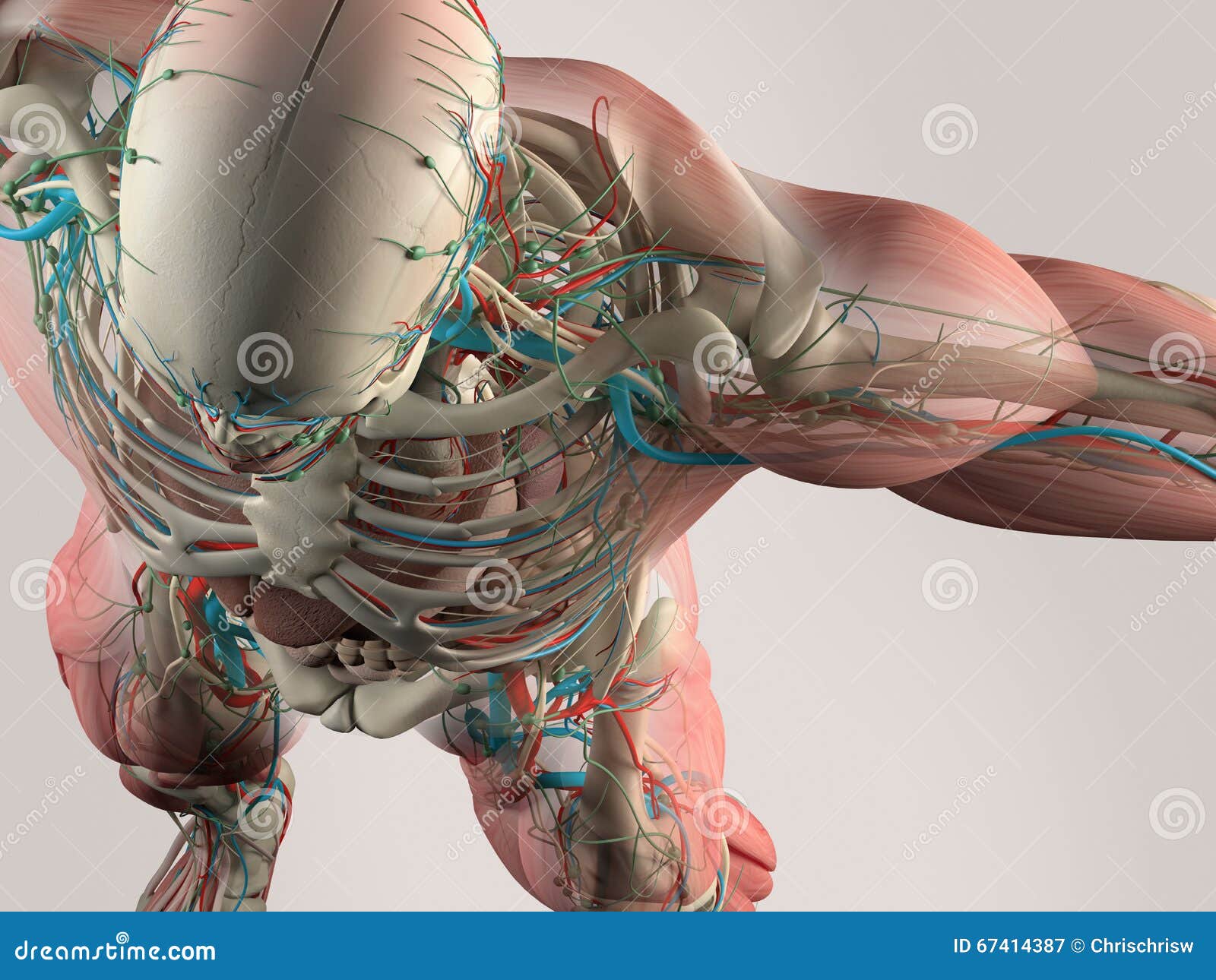
Human Anatomy Detail Of Chest And Shoulder Muscle Arteries On Plain Studio Background Human Anatomy Detail Of Skull And Shoulde Stock Illustration Illustration Of Male Muscles 67414387 from thumbs.dreamstime.com These myotomes divide into the epimere and the hypomere. A good radiologist knows the anatomy because knowing where structures normally live and recognizing the location of an abnormality helps to make or narrow the differential diagnosis. The muscles of the chest develop from the somites found in the mesoderm. Learn about each of these muscles, their locations, functional anatomy and exercises for them. In insects, crustaceans, and the extinct trilobites, the thorax is one of the three main divisions of the creature's body, each of which is in turn composed of multiple segments. The first step in understanding thorax anatomy is to find out its boundaries. The thorax has two major openings: Anatomy of the chest, abdomen, and pelvis was produced in part due to the generous funding of the david f.
These myotomes divide into the epimere and the hypomere.
Sternocleidomastoid muscle clavicle and ribs anatomy muscle anatomy chest sternocleidomastoid ribs anatomy chest muscles anatomy thorax rib muscles chest muscles chest anatomy illustration. A line is drawn from anterior surface of the body of 6th thoracic vertebrae passing through the apex of the heart up to anterior lower most part of diaphragm. About the 6th week, the somites differentiate into the sclerotomes and the dermatomyotomes. Radiology basics of chest ct anatomy with annotated coronal images and scrollable axial images to help medical students and junior doctors learning anatomy. Computed tomography (ct) of the chest can detect pathology that may not show up on a conventional chest radiograph(1). It is a flat bone that articulates with the clavicle and the costal cartilages of the upper 7 ribs (true ribs), while the 8th,. Anatomy of the thorax, heart, abdomen and pelvis recommended text gray's anatomy for students. The chest is made up primarily of two muscles: Anatomy of the chest and shoulder, anatomy of the chest organs, anatomy of the chest wall, anatomy of the chest wall and pleura, anatomy of upper chest area, human. The epidermis is the outermost layer that provides a protective, waterproof seal over the body. Muscles of the chest and their functions you have two mighty muscles on both sides of your chest: It provides protection to vital organs (eg, heart and major vessels, lungs, liver) and provides stability for movement. The thorax has two major openings:
System respiratory respiratory organs of human body digestive and respiratory system medical chest internal structure of human body medicine body lungs biology intestines stomach anatomy torso human internal. Computed tomography (ct) of the chest can detect pathology that may not show up on a conventional chest radiograph(1). Plus, how to target each to make them bigger and stronger. Chest a man's chest — like the rest of his body — is covered with skin that has two layers. The chest is the area of origin for many of the body's systems as it houses organs such as the heart, esophagus, trachea, lungs, and thoracic diaphragm.
1 Anatomy Of The Arm And Chest Used With Permission Of N Moureau Download Scientific Diagram from www.researchgate.net See human chest anatomy stock video clips. Anatomy of the thorax, heart, abdomen and pelvis recommended text gray's anatomy for students. The muscles of the chest develop from the somites found in the mesoderm. Angina is the term for chest pain caused by poor blood flow to the heart. It is important to remember the position and orientation of the heart when placing a stethoscope on the chest of a patient and listening for heart sounds, and also when looking at images taken from a midsagittal perspective. The epidermis is the outermost layer that provides a protective, waterproof seal over the body. Anatomy of the chest and shoulder, anatomy of the chest organs, anatomy of the chest wall, anatomy of the chest wall and pleura, anatomy of upper chest area, human. The chest or thorax is the region between the neck and diaphragm that encloses organs, such as the heart, lungs, esophagus, trachea, and thoracic diaphragm.
Normal anatomy of the thorax on labeled chest ct:
Angina is the term for chest pain caused by poor blood flow to the heart. It is enclosed by the ribs, the vertebral column, and the sternum, or breastbone, and is separated from the abdominal cavity (the body's largest hollow space) by a muscular and membranous partition, the diaphragm. Anatomy of the thorax, heart, abdomen and pelvis recommended text gray's anatomy for students. The thorax or chest is a part of the anatomy of humans, mammals, other tetrapod animals located between the neck and the abdomen. Plus, how to target each to make them bigger and stronger. The chest is made up primarily of two muscles: The chest or thorax is the region between the neck and diaphragm that encloses organs, such as the heart, lungs, esophagus, trachea, and thoracic diaphragm. The human thorax includes the thoracic cavity and the thoracic wall. Learn about each of these muscles, their locations, functional anatomy and exercises for them. The right side of the heart is deflected anteriorly, and the left side is deflected posteriorly. The first step in understanding thorax anatomy is to find out its boundaries. Chest pain has many possible causes, all of which need medical attention. Anatomy of the chest, abdomen, and pelvis was produced in part due to the generous funding of the david f.
A heart attack results from blocked blood flow, often from a blood clot, to your heart muscle. Download my two educational text books for free using this link: The chest or thorax is the region between the neck and diaphragm that encloses organs, such as the heart, lungs, esophagus, trachea, and thoracic diaphragm. The chest is made up primarily of two muscles: Anatomy of the thorax, heart, abdomen and pelvis recommended text gray's anatomy for students.
Human Chest Anatomy Images Stock Photos Vectors Shutterstock from image.shutterstock.com Angina is the term for chest pain caused by poor blood flow to the heart. Muscles of the chest and their functions you have two mighty muscles on both sides of your chest: The chest anatomy includes the pectoralis major, pectoralis minor and the serratus anterior. The chest or thorax is the region between the neck and diaphragm that encloses organs, such as the heart, lungs, esophagus, trachea, and thoracic diaphragm. The thorax has two major openings: The thorax or chest is a part of the anatomy of humans, mammals, other tetrapod animals located between the neck and the abdomen. It is a flat bone that articulates with the clavicle and the costal cartilages of the upper 7 ribs (true ribs), while the 8th,. Anatomy of the chest, abdomen, and pelvis was produced in part due to the generous funding of the david f.
The epidermis is the outermost layer that provides a protective, waterproof seal over the body.
Anatomy of pancreas 12 photos of the anatomy of pancreas anatomy and histology of pancreas ppt, anatomy and physiology of human pancreas, anatomy of pancreas divisum, describe the microscopic anatomy of pancreas, gross anatomy of pancreas, human anatomy, anatomy and histology of pancreas ppt, anatomy and physiology of. Browse 6,407 chest anatomy stock photos and images available, or search for human anatomy to find more great stock photos and pictures. Swensen fund for innovation in teaching. Radiological anatomy of the lungs, mediastinal lymph nodes, trachea, bronchi, pleural cavity, heart and pulmonary vessels. The epidermis is the outermost layer that provides a protective, waterproof seal over the body. The chest is the area of origin for many of the body's systems as it houses organs such as the heart, esophagus, trachea, lungs, and thoracic diaphragm. An overview of the anatomy visible in a transverse computed axial tomographical image of the thorax (and part of the abdomen) performed with intravenous cont. The myotomes elongate and invade the mesoderm of the wall of the embryonic thoracic and abdominal cavities. The chest wall is comprised of skin, fat, muscles, and the thoracic skeleton. This chapter is an abbreviated review of thoracic anatomy as seen on chest radiographs and computed tomography (ct) of the chest. A heart attack results from blocked blood flow, often from a blood clot, to your heart muscle. The chest anatomy includes the pectoralis major, pectoralis minor and the serratus anterior. It is enclosed by the ribs, the vertebral column, and the sternum, or breastbone, and is separated from the abdominal cavity (the body's largest hollow space) by a muscular and membranous partition, the diaphragm.